Evidence-based diagnosis of lower back pain.
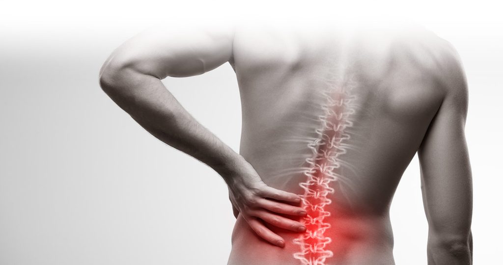
Lower back pain (LBP) refers to discomfort or pain in the lumbar region of the spine, which is the area below the ribcage and above the pelvis. Dr Tony Kostos, Rheumatologist, discusses methods of gaining evidence and the best approach.
How do we ascertain the history of Lower Back Pain?
No features in the lower back pain history can reliably determine the cause. Instead, we can approach history to determine if there are any psychosocial factors or more serious conditions that warrant further investigations.
Psychosocial factors known as yellow flags can include:
- Attitudes and beliefs about Lower Back Pain.
- Diagnostic and treatment issues.
- Compensation issues.
- Behaviour and emotions.
- Work
- Family.
Serious factors known as red flags can include:
- Inflammatory Disease: For example, a 26-year-old female experiencing pain at night with EMS and immobility stiffness. Her pain can improve with an exercise regime.
- Malignancy: This on the other hand as an example is a person over the age of 50 with previous experience of malignancy. They have had prolonged illness especially with weight loss and fails to improve even with treatment.
- Infection: Someone experiencing nocturnal pain with systemic symptoms and body penetration.
- Fractures: In this instance, it’s due to trauma to the body. Minor trauma, Osteoporosis or Corticosteroid use are most common causes.
The examination process
Medical experts can establish if the spine is stiff and whether features of abnormal illness behaviour and non-organic signs are present. However, physical examination cannot provide a diagnosis of LBP, as commonly performed tests lack reliability, specificity, or both.
As we established, history and examination alone cannot determine the cause of LBP. Using investigation methods such as X-rays, scintigraphy, and CT scans cannot determine the cause of LBP either. MRI scans, however, are the best screening test for red flag conditions. MRI helps identify disc degeneration, disc herniation, and bulges that can all occur in asymptomatic individuals with increasing frequency with increasing age. Please note that MRI scans don’t predict the future.
“The findings on MRI were not predictive of the development or duration of LBP. Individuals with LBP did not have the greatest degree anatomical abnormality on the original 1989 scans.”
Borenstein et al. JBJS AM 2001;83-A:1306-11
Other methods of investigations
Following MRI scans, Medial branch blocks (MBB) is a test to help the doctor understand whether pain arising from the patient’s facet joints can be relieved by numbing the nerves During a lumbar medial branch block, the pain intervention specialist will deliver a long-acting local anaesthetic to the medial branch nerves that supply the facet joints in your low back. This procedure is performed with strict methods to obtain meaningful results. Forthwith goes the same for sacroiliac blocks which is primarily used either to diagnose or treat low back pain and/or sciatica symptoms associated with sacroiliac joint dysfunction.
Additional examinations include high-intensity zones (HIZs) that may indicate that the disc is painful but can be seen in asymptomatic individuals. If a discography (another imaging test used to evaluate back pain) takes place, it needs to be performed using strict criteria, and results need to be interpreted with caution.
The multifaceted nature of lower back pain necessitates a thorough and careful approach to history, examination, and specialised investigations. This is in order to unravel its complex aetiology and guide effective management strategies.
Australian Specialist Hub provides Rheumatology experts and specialists in all other medical areas. We also have an extensive panel of Occupational Therapists across all states. Our panel of experts delivers evidence-based quality reports in a timely, cost-effective manner.
Please call our friendly team at 1800 287 482 for further assistance or submit your referral, and we will get back to you shortly.


